Most patients arrive at their first implant consultation expecting a conversation about the implant. What they often get instead is a conversation about bone. When a dentist recommends a bone graft before dental implants, the finding underneath it is simple: the alveolar ridge is no longer thick or tall enough to hold a fixture securely.
This is one of the most common results of a first consultation for international patients, and the single most common reason a treatment timeline turns out longer than the one they planned their flights around. Dental bone grafting, also called alveolar bone grafting, rebuilds the missing bone at the implant site so the fixture has a foundation capable of holding it for years rather than months.
The question that matters to most patients is not what a graft is. It is whether they need one, and what it does to their schedule. This guide answers both.
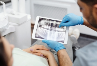
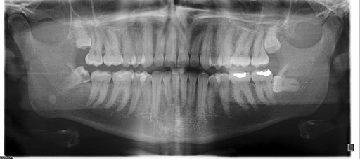
Reduced bone volume gives almost no warning. It is not painful, it is rarely visible in the mirror, and most patients learn about it only when 3D imaging goes up on the screen at a consultation.
Certain histories make the finding considerably more likely:
None of these confirm that a graft is required. What they establish is that bone height, bone width, and bone quality all need to be measured properly before an implant plan is finalized, rather than assumed.
The underlying cause is straightforward. The alveolar bone stays healthy largely through the pressure of chewing, and once a tooth is gone that stimulation stops. Resorption follows on its own, fastest in the months immediately after extraction. The companion guide on when to get full mouth implants after extractions covers that timeline in detail.
An implant does not simply sit in the jaw. It transmits chewing force into the bone around it, which means the bone has to be capable of receiving that force on every side of the fixture. Where the remaining ridge is too thin or too shallow, that support is missing, and the implant starts at a disadvantage from the first day it is loaded.
Grafting restores both the width and the height of the ridge. It can also be performed at the moment of extraction, filling the socket to limit the shrinkage that would otherwise take place over the following months.
This is worth stating plainly, because patients frequently read a graft recommendation as an upsell. For many cases it is the opposite. Grafting is not an addition to implant treatment. It is often the thing that makes implant treatment possible at all.
Three procedures cover the large majority of cases. Which one applies depends on where the bone is missing and how much of it is gone.
Performed immediately after an extraction, socket preservation places graft material into the empty socket and covers it with a collagen membrane or a similar barrier. The purpose is to limit the natural shrinkage of the ridge and keep the site in better condition for an implant later.
For patients who already know an implant is planned, this one step at the time of extraction can remove the need for a much larger graft procedure months down the line. It is the cheapest and least invasive point in the entire process to intervene, and the one most often missed.
The upper molar area sits directly beneath the maxillary sinus, and after tooth loss the sinus tends to expand downward into the space. Where the remaining bone height is insufficient, a sinus lift raises the sinus membrane carefully and places graft material beneath it, creating the vertical support the implant requires.
Where bone height is inadequate and this step is skipped, the implant may be left without stable support, and the sinus area itself can be affected.
Guided bone regeneration, usually shortened to GBR, applies where the shape of the ridge has partially collapsed. Gum tissue regenerates faster than bone does, so left alone it fills the defect before bone cells can. A membrane holds that space open and keeps the soft tissue out, allowing bone to rebuild in a protected environment.
For patients traveling to Korea, this is the section that determines the trip.
Same-day placement puts the graft and the implant in during a single surgery. It becomes an option when enough native bone remains to achieve initial stability for the fixture at the time of placement.
Staged grafting separates the two. The graft goes in first and is left to mature for roughly three to six months. The implant follows once the foundation has consolidated.
The decision rests on bone volume, bone quality, infection risk, and the specific site. It is a clinical judgment rather than a scheduling preference, and it cannot be settled before imaging has been reviewed. A patient who books a two-week trip on the assumption of same-day treatment, and then learns at the consultation that staging is the safer route, has a problem that no amount of clinic flexibility can solve.
International patients usually plan a single trip. Where a graft is required, the realistic structure is two.
First visit. Diagnosis, 3D imaging, any extraction, and the graft. This generally requires around one to two weeks in Korea, depending on how early healing progresses.
Healing interval. Three to six months, spent at home. Nothing about this period requires the patient to be in Korea.
Second visit. Implant placement and the prosthetic phase.
Same-day treatment compresses this considerably, and for suitable cases it is genuinely a single trip. The practical difficulty is that suitability cannot be confirmed remotely without records.
This is why a remote review of existing dental records and imaging is worth arranging before flights are booked rather than after. It allows a provisional plan, a realistic number of visits, and an approximate length of stay to be discussed while those things can still be changed.
Four categories of material are in routine use, and the choice depends on the patient's condition, the size of the defect, and the site being treated.
Autogenous bone is taken from the patient's own body. Biological compatibility is very high, since the tissue is recognised as the patient's own, though harvesting it requires a second surgical site.
Allograft bone comes from processed human donor bone and supports regeneration without that second site.
Xenograft bone is generally derived from bovine or porcine sources. The organic component is removed, leaving a mineral scaffold for new bone to grow into.
Synthetic bone is manufactured from biocompatible materials such as calcium phosphate and holds space open while new bone forms.
In practice, clinics in Korea frequently combine them, pairing a scaffold material that maintains volume with a more biologically active one that encourages regeneration. Modern graft materials are generally predictable when selected and handled appropriately, though the result still depends on the patient's overall health, the condition of the surgical site, and the treatment plan itself.
Swelling and mild discomfort are usual for several days, with the first 48 hours typically the most noticeable. Soft foods are generally recommended during early healing, and chewing on the treated side is usually avoided.
Smoking matters more here than patients tend to expect. Nicotine constricts blood vessels and reduces blood flow to the surgical site, which directly interferes with bone formation. Alcohol is generally avoided during early healing for related reasons. Taking prescribed medication as directed and keeping the area clean both have a measurable effect on how well the graft integrates.
Grafted bone generally requires about three to six months to mature before it can support an implant, with the range depending on the size of the defect and the patient's general health.
That the CT scan settles it. Imaging carries a great deal of weight, but bone quality, soft tissue condition, and the surgeon's assessment during planning and sometimes during the procedure itself all feed into the decision. A scan narrows the question rather than closing it.
That a graft makes the surgery much more painful. Discomfort afterwards tends to track incision size, length of surgery, and how the tissue was handled, rather than the presence of graft material.
That graft materials are risky. Bone grafting performed in a sterile environment with verified materials is generally safe. Adherence to aftercare is what keeps infection risk low.
That a shorter implant avoids the whole problem. Shorter and narrower fixtures are appropriate in selected cases, but they are not a general substitute for adequate bone support, and placing them where support is inadequate may raise the risk of failure.
How can a patient tell whether bone grafting is needed? Not reliably, without imaging. Reduced bone volume is common among patients missing a tooth for a long period, patients with gum recession, and patients who lost teeth to periodontal disease, but confirmation requires a clinical examination combined with 3D imaging of bone height and width.
Is one graft material better than the others? No single material suits every patient. The choice depends on general health, the size of the defect, and the area being treated, and materials are frequently combined to support regeneration while maintaining space.
Why is a sinus lift needed for upper molar implants? The upper molar area sits close to the maxillary sinus, and after tooth loss the sinus can expand downward into the available space. Where bone height is insufficient, a sinus lift creates the vertical support the implant needs and helps protect the sinus during placement.
What should patients avoid after bone grafting? Smoking and alcohol, particularly during early healing. Nicotine reduces blood flow to the site and can interfere with bone formation. Prescribed medication taken as directed and careful cleaning of the area are equally important.
How long does healing take after dental bone grafting? Grafted bone generally requires about three to six months to mature. Where grafting and implant placement are performed together, the overall timeline can be shorter, though this depends on the initial condition of the bone.
Can bone grafting and implants be completed during one trip to Korea? Sometimes, where enough native bone remains for one-stage treatment. Where bone loss is significant, two visits separated by several months of healing is generally the safer structure. This can only be determined once imaging has been reviewed.
Bone grafting rebuilds the support that stable implant treatment depends on. With accurate diagnosis, verified materials, and a plan built around the individual site, it makes implant treatment considerably more predictable than placing a fixture into bone that cannot hold it.
For patients considering implant treatment in Korea, the practical first step is a remote review of existing dental records and imaging. It allows a provisional plan and timeline to be discussed before flights, accommodation, and time off work are committed to. KORA coordinates that review with its partner dental clinics, arranges the consultation, and stays with the patient through treatment and recovery.
This article was written by KORA based on an interview with Dr. Jin Hwan Kim, Director of Oneday Dental Clinic.
It provides general information only and is not a diagnosis or a treatment recommendation. Suitability, technique, and healing timelines vary by individual and are determined by the treating dentist following clinical examination and 3D imaging.